Patients and Parents
 Visitors
 Healthcare Professionals
 Research
 Donate and Support
 About HKCH
HKCH Newsletter
Neuroimaging: decoding the maze of the brain
- Share :
-

-

-

-


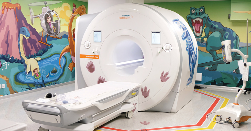
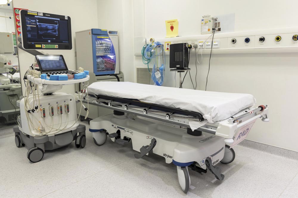
The central nervous system governs crucial bodily functions, but children often struggle to articulate their symptoms when abnormalities arise. In these critical moments, neuroimaging plays a vital role in showing what’s wrong in the brain and spine, solving complex medical mysteries.
Neuroimaging covers a vast spectrum, ranging from neurosurgical conditions such as brain tumors, to neurological disorders affecting the central nervous system. It also captures brain and spinal lesions caused by metabolic diseases. These complexities, coupled with patients' critical conditions, make diagnosis challenging.
Dr Chris Wong, Consultant (Radiology) explained, "Some neurological conditions are rare and come in many forms, showing very different symptoms across age groups. We don't just interpret images. We must delve deep into the underlying pathology and genetic mutations to deliver an accurate diagnosis."
Real-time precision navigation for surgeries
For complex surgeries, the radiology team may perform real-time intraoperative imaging to provide precise navigation for the surgeons.
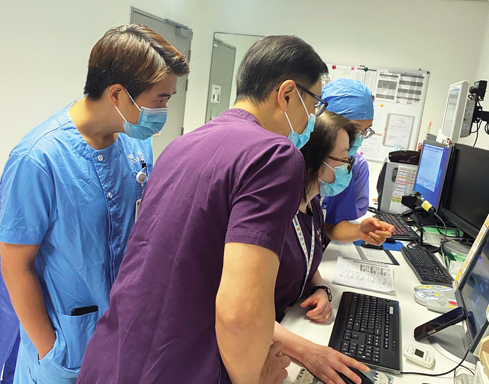
There was a child who suffered from frequent seizures caused by a hypothalamic hamartoma in the brain. HKCH Neurosurgery, Radiology, Anaesthesiology, and Queen Mary Hospital's Neurosurgery collaborated to perform a stereotactic radiofrequency thermocoagulation, successfully blocking the abnormal signals between the lesion and the brain. This significantly reduced the child's seizure frequency, marking the first paediatric case of its kind in Hong Kong.
Prior to the surgery, radiologists and neurosurgeons carefully analyzed the patient's MRI images, mapping the lesion, neural structures and blood vessels for surgical planning.
Dr Wong recalled, "On the day of surgery, once the child's head was secured in a stereotactic frame at the operating theatre, we immediately performed CT scan with metallic artefacts reduction technique in our department. Neurosurgeons then fused this CT image with the pre-operative MRI image, clearly visualizing the head frame, brain structures and lesion to precisely implant the electrodes into the targeted location in the brain at the operating theatre. Once the placement was confirmed with a second CT scan, radiofrequency was applied to successfully destroy the lesion signal."