Patients and Parents
 Visitors
 Healthcare Professionals
 Research
 Donate and Support
 About HKCH
HKCH Newsletter
Interventional radiology: navigating hope
- Share :
-

-

-

-


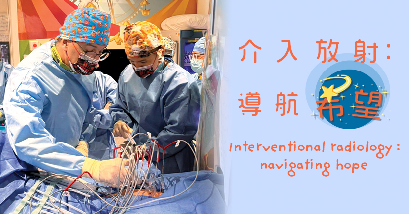
When people think of radiology, they often associate it with imaging exams. In fact, the HKCH Department of Radiology also provides an important service that combines image guidance with minimally invasive procedures —interventional radiology (IR).
Dr Kevin Fung, Consultant (Radiology) explained that IR uses real-time ultrasound, CT, MRI and X-ray to guide catheters or needles precisely to the lesion for diagnosis and treatment. "Instead of opening up the body to perform surgery, we use medical imaging to guide treatment." With small incisions, IR reduces trauma and pain while promoting faster recovery.
At HKCH, the IR team provides image guided minimally invasive treatments for a wide range of complex conditions, including tumours, renovascular hypertension, vascular malformations and lymphatic disorders.
Innovative solutions for complex cases
The value of paediatric IR is best demonstrated in the most challenging cases. A two-week-old premature baby developed heart failure caused by a massive congenital hepatic haemangioma. The team planned an embolization to block the tumour's blood supply.
Dr Fung recalled, "The baby's total blood volume was only about 200 ml – roughly a mini can of cola. Every step had to be calculated with utmost precision. The biggest challenge was the extremely tiny blood vessels, making a conventional femoral approach risky for thrombosis."
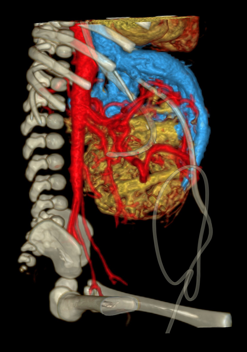
To win the race against time, the team adopted an innovative approach – entering through the baby's umbilical cord, then advanced through her patent ductus arteriosus (which was still patent shortly after birth) near the heart, reaching the aorta and finally the tumour. The embolization was completed successfully.
This breakthrough method was published in an international medical journal. Dr Fung was selected to present the case at a leading international IR conference, underscoring HKCH's IR services have reached international standards.